Western Blotting Starter Kit 2 (TBS/PVDF)
Components
These are the components for the Western Blotting Starter Kit 2 (TBS/PVDF), PN 925-35011:
P/N |
Description |
926-68170 |
IRDye® 680RD Goat anti-Mouse Secondary Antibody, 25 µL, 1 mg/mL |
827-08365 |
IRDye 800CW Goat anti-Rabbit Secondary Antibody, 25 µL, 1 mg/mL |
927-60000 |
Intercept® (TBS) Blocking Buffer, 125 mL |
827-16547 |
Immobilon®-FL PVDF Membranes, 4 (10 x 10 cm) |
Quick Start Guidelines for Western Blot Preparation
-
Blots can be prepared in advance and stored.
- Prepare replicate blots when comparing established methods.
- One following your current protocol.
- One following the LI‑COR protocol for detection with an Odyssey Imaging System.
- Optional: Prepare a blot using current methods and reagents but detect with secondary antibodies from LI‑COR.
-
Standard pre-stained blue molecular weight markers can be visualized on Odyssey Imaging Systems.
Because of the sensitivity of Odyssey Imagers, load only 25% of the normal volume. For best results, leave the markers barely visible on the gel, otherwise the marker will be too strong on the Odyssey image.
-
Do not write on membranes with regular ink pens or markers, because the ink will fluoresce on Odyssey Imaging Systems. Use a pencil to write on the membrane.
-
Only handle membranes by the edges with clean forceps. Be careful not to touch the membrane with your hands or gloves.
-
For one color blots, use IRDye 800CW secondary antibody for detection of the protein.
- For two color blots:
- Ensure primary antibodies are from different species (e.g., rabbit polyclonal and mouse monoclonal).
- Ideally, use the IRDye 800CW secondary antibody to detect the low abundant protein and IRDye 680RD to detect the more abundant protein.
-
If you normally use PVDF membranes, use the PVDF membrane included in this kit.
Nitrocellulose can also be imaged on the Odyssey Imager.
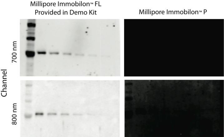
Comparison of Millipore Immobilon-FL PVDF and Immobilon-P Western blots on the Odyssey Classic Imaging System.
-
When processing Western blots, do not use any dishes that have been used for Coomassie staining. Odyssey Imagers are very sensitive to Coomassie and dishes used for staining will cause high background in the 700 channel.
-
Do not add Tween® 20 to Intercept Blocking Buffer during the blocking step.
-
Dilute your primary antibody in Intercept Blocking Buffer to the same concentration as you use in your current detection method. Add 0.2% Tween 20 to the final concentration. Incubate the usual amount of time.
Do not reduce the concentration of your primary antibody.
Western Blot Detection Protocol
Prepare your Western blot using standard blotting procedures and the membrane provided in this kit. After transfer, allow the blot to dry for an hour before beginning the detection protocol. Dry blots can be stored between pieces of filter paper overnight at room temperature.
Only handle membranes by the edges with clean forceps. Be careful not to touch the membrane with your hands or gloves.
Do not write on membranes with regular ink pens or markers, because the ink will fluoresce on Odyssey Imaging Systems. Use a pencil to write on the membrane.
Step 1. Wet Membrane
For Immobilon®-FL PVDF Membranes
- Wet for 30 seconds in 100% methanol.
- Wet in 1X
Step 2. Block the Membrane
Place the membrane in an incubation box and block the membrane with Intercept® Blocking Buffer for 1 hour at room temperature with gentle shaking.
Be sure to use sufficient blocking buffer to cover the membrane (a minimum of 0.4 mL/cm2 is suggested).
Step 3. Dilute Primary Antibody
-
Use Intercept T20 Antibody Diluent as the primary antibody diluent or prepare your own by adding Tween® 20 to Intercept Blocking Buffer for a final concentration of 0.2% Tween 20.
-
Dilute primary antibody in antibody diluent using the vendor's recommendations.
- Depending on the primary antibody, dilutions may range from 1:200 – 1:5,000.
- Use enough antibody solution to completely cover the membrane.
Step 4. Incubate Blot in Diluted Primary Antibody
Incubate the blot for 1 - 4 hours at room temperature or overnight at 4 °C with gentle shaking.
Optimal incubation times vary for different primary antibodies.
If the procedure cannot be completed in full, this is a good place to stop until the following day. Incubate the primary antibody overnight at 4 °C with gentle shaking.
Step 5. Wash Membrane
- Carefully pour off primary antibody solution.
- Rinse the membrane with 1X TBS-T (0.2% Tween 20) or 1X PBS-T (0.2% Tween 20).
- Cover blot with 1X TBS-T or 1X PBS-T.
- Shake vigorously on platform shaker at room temperature for 5 minutes.
- Pour off wash solution.
- Repeat steps 3 to 5 for a total of 4 washes.
Step 6. Dilute Secondary Antibody
Dilute secondary antibody in the appropriate diluent for the membrane you're using. For IRDye® 800CW secondary antibodies, IRDye® 680RD secondary antibodies, or IRDye® 680LT secondary antibodies, the recommended starting dilution is 1:20,000. For VRDye secondary antibodies, the recommended starting dilution is 1:10,000. The following includes recommendations on antibody dilution buffers.
Secondary Antibody Diluent for Immobilon®-FL PVDF
Use Intercept T20 Antibody Diluent and SDS. Alternatively, add Tween 20 to a final concentration of 0.2% and SDS to a final concentration of 0.01 - 0.02% in Intercept Blocking Buffer.
Step 7. Incubate Blot in Secondary Antibody
Protect membrane from light during incubation.
- Incubate blot in diluted secondary antibody for 1 hour at room temperature with gentle shaking.
- Do not incubate for longer than 1 hour, because the background may increase.
Step 8. Wash Membrane
Protect membrane from light during washes.
- Carefully pour off secondary antibody solution.
- Rinse the membrane with 1X TBS-T or 1X PBS-T.
- Cover blot with 1X TBS-T or 1X PBS-T.
- Shake vigorously on platform shaker at room temperature for 5 minutes.
- Pour off wash solution.
- Repeat steps 3 to 5 for a total of 4 washes.
Step 9. Rinse Membrane
Rinse the membrane with 1X
-
Membranes can be stored at 4 °C in TBS or PBS for short periods. Always protect membranes from light.
-
Membranes can be stored dry at room temperature for prolonged storage. Always protect membranes from light.
- If you plan to strip and reprobe the Western blot, do not allow the completed Western blot to dry. The stripping process is less effective on Western blots that have been allowed to dry.
Step 10. Scan Membrane
Protect the membrane from light prior to scanning.
Scan the membrane on an Odyssey Imager.
The membrane can be scanned wet or dry. Scanning the membrane dry can add signal intensity, but can also lead to increased background.
Optimization
-
Follow the protocol carefully.
-
No single blocking buffer will be optimal for every antigen-antibody pair.
Some primary antibodies may exhibit greatly reduced signal or non-specific binding in different blocking solutions. If you have difficulty detecting your target protein, changing the blocking solution may dramatically improve performance. If the primary antibody has worked well in the past using chemiluminescent detection, try that same blocking solution for near-infrared fluorescent detection on an Odyssey Imaging System.
-
To avoid background speckles on blots, use high-quality ultra pure water for buffers. Rinsing previously-used incubation boxes with methanol can reduce background contamination of future blots.
-
Always use a consistent buffering system for wash buffers and blocking buffers. (e.g. Intercept (TBS) Blocking Buffer, use TBS as the base for wash buffers; for Intercept (PBS) Blocking Buffer, use PBS as the base for wash buffers).
-
Never perform Western incubations or washes in dishes that have been used for Coomassie staining.
-
Only handle membranes by the edges with clean forceps. Be careful not to touch the membrane with your hands or gloves.
After handling membranes that have been incubating in antibody solutions, clean forceps thoroughly with methanol, then rinse with distilled water.
-
Always pour off antibody solution and washes from the same corner of the box to ensure complete removal of previous solutions.
-
Do not wrap the membrane in plastic when scanning.
Guidelines for Multiplexed Detection
Multiple antigens can be detected simultaneously on the same blot using IRDye® secondary antibodies and VRDye™ secondary antibodies. When performing a blot, use the standard Western blot protocol with the following modifications:
-
Combine the primary antibodies in the antibody diluent. Incubate simultaneously with the membrane. The primary antibodies must be from different host species or subclasses if the targets are close in molecular weight.
-
Combine the IRDye secondary antibodies and VRDye secondary antibodies in the antibody diluent.
-
Incubate simultaneously with the membrane.
Multiplexed detection requires careful selection of primary and secondary antibodies. A comprehensive course is available on the Lambda U® Western Blot Education portal (lambdau.net) to help you select appropriate antibodies. The following guidelines provide information that will help you successfully design multiplexed experiments:
-
The primary antibodies must be derived from different host species so that they can be discriminated by secondary antibodies of different specificities (for example, primaries from rabbit and mouse will be discriminated by anti-rabbit and anti-mouse secondary antibodies, respectively).
-
If the two primary antibodies are mouse monoclonals from different IgG subclasses (e.g., IgG1, IgG2a, or IgG2b), IRDye® Subclass-Specific secondary antibodies can be used for multiplex detection. The same subclasses cannot be combined in a two-color Western blot (e.g., two IgG1 primary antibodies).
See Western Blot and In-Cell Western™ Assay Detection Using IRDye Subclass-Specific Antibodies (licorbio.com/subclass) for more information.
-
Anti-goat secondary antibodies cannot be multiplexed with goat-derived secondary antibodies (e.g., donkey anti-goat and goat anti-rabbit). The secondary antibodies will cross-react.
-
Before combining primary antibodies in a multiplexed experiment, always perform preliminary blots with each primary antibody alone to determine the expected banding pattern and possible background bands. Slight cross-reactivity may occur and can complicate interpretation of your blot, particularly if the antigen is very abundant. If cross-reactivity is a problem, load less protein or reduce the amount of antibody.
-
One secondary antibody must be labeled with IRDye 680RD secondary antibody, IRDye 680LT secondary antibody, or VRDye secondary antibodies. The other must be labeled with IRDye 800CW secondary antibody or VRDye secondary antibodies.
-
It is generally recommended that the IRDye 800CW secondary antibody (800 nm channel) be used to detect the lower-abundance protein target and IRDye 680RD secondary antibody (700 nm) to detect the more abundant protein. If using an instrument that can also detect visible fluorescence, use those channels to detect the internal loading control using Revert 520 Total Protein Stain (preferred) or VRDye 490 Secondary Antibodies in the 490 nm channel or VRDye 549 Secondary Antibodies in the 520 nm channel.
-
Always use highly cross-adsorbed secondary antibodies for multiplexed detection. Failure to use cross-adsorbed antibodies may result in increased cross-reactivity.
-
For best results, avoid using primary antibodies from mouse and rat together in a multiplexed experiment. The two species are so closely related it is not possible to completely adsorb away all cross-reactivity. If there is no other option but to use mouse and rat together, it is crucial to run single-color blots first with each individual antibody to be certain of expected band sizes.
-
If possible, the secondary antibodies should be derived from the same host species (e.g., donkey anti-mouse and donkey anti-goat) to eliminate the chance of the secondary antibodies reacting against one another.
-
High abundant targets, such as housekeeping proteins, may perform better in the 490 or 520 channels. Consequently, their working range can be identified and combined with lower abundant targets in the 700 and 800 channels using VRDye secondary antibodies.