Protein Array

Protein arrays are a high-throughput way to generate information about protein abundance or modification state.

When you use near-infrared fluorescence to detect your protein arrays, you will get:

  • Dramatically reduced background autofluorescence of nitrocellulose-coated slides 1
  • Sensitivity in the femtogram range 2
  • Wide dynamic range
  • Ability for multiplexed detection to quantify and compare two targets 3
  • Simplified detection protocol 3

Common Types of Protein Arrays 4

Protein arrays with spots that are equal to or greater than 85 μm are compatible with the Odyssey® DLx Imager, whereas spots that are equal to or greater than 500 μm are compatible with the Odyssey XF Imager.
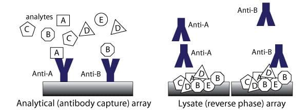
  • Lysate (reverse phase) arrays contain complex samples, such as cell or tissue lysates, that are printed on an array surface and interrogated with antibodies.
  • Analytical arrays use affinity reagents such as antibodies or peptides to profile analytes in complex mixtures of proteins. These arrays can be spotted on a chip or slide, or spotted into microwell plates. Antibody capture arrays are the most common form. Examples of analytical arrays are the Quansys ELISA arrays.
  • Functional protein arrays are spotted with many different purified proteins, and used to assay biochemical functions such as protein-protein, protein-DNA, and protein-small molecule interactions and enzyme activity.
figure 1
Figure 1. Schematic of lysate and analytical protein array concepts. Adapted from Sheehan, KM et al.
figure 2
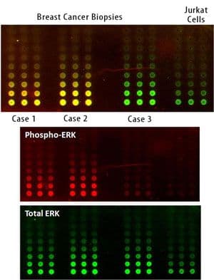
Figure 2. Multiplexed detection of phosphorylated and total ERK protein levels. Tissue samples were obtained for three human breast cancer patients; lysates were prepared from these and from a Jurkat T cell control. The array was probed simultaneously with antibodies against phospho-ERK (mouse) and total ERK (rabbit), and detected with near-infrared labeled secondary antibodies (red, green). Overlaid image of total and phospho-ERK levels is shown at the top (red + green = yellow). Adapted from Calvert, VS et al.

Reverse Phase (Lysate) Arrays

Lysate (reverse phase) arrays contain complex samples, such as cell or tissue lysates, that are printed on an array surface and interrogated with antibodies.4
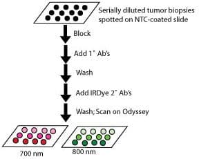
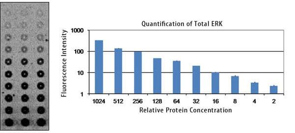
figure 3
Figure 3. Schematic protocol for near-infrared lysate (reverse phase) arrays. Adapted from Calvert, VS et al.

Advantages of Lysate Arrays

  • More quantitative and replicable than Westerns (intra-chip variation ˜0.1%) 2
  • Wide dynamic range
  • Conservation of precious samples
  • Very sensitive — can detect femtogram or single-cell protein levels 2
  • Absolute quantification when spotting recombinant protein standards 2
  • Process many replicates and dilutions easily
figure 4
Figure 4. Replicability of ERK detection using infrared dyes. Lysate was prepared from a human breast cancer tissue sample. It was arrayed in triplicate in a series of two-fold dilutions, and stained with rabbit anti-ERK primary Ab and IRDye® 800CW dye-labeled goat anti-rabbit. Linearity and replicability of this data is shown in the graph. The average of three replicates is plotted; error bars are included but are very small and difficult to see. Adapted from Calvert, VS et al.

References

  1. Sheehan, KM et al. (2005). Molecular & Cellular Proteomics, 4(4), pp 346-55. DOI: 10.1074/mcp.T500003-MCP200.
  2. Loebke, C et al. (2007). Proteomics, 7(4), pp 558-64. DOI: 10.1002/pmic.200600757.
  3. Ambroz, K et al. (2005). Gene Expression Analysis Using Infrared Fluorescent Labeling and Detection Methods. Poster presented at Chips to Hit Annual Meeting.
  4. Hall, DA et al. (2007). Mechanisms of Ageing and Development, 128(1), pp 161-67. DOI: 10.1016/j.mad.2006.11.021.
  5. Calvert, VS et al. (2004). Clinical Proteomics, 1(1), pp 81-89. DOI: 10.1385/CP:1:1:081.